+86 13683842418    
625006192@qq.com
Menu
What are you looking for?
  • +86 13683842418

  • +86 13683842418

  • 河南省洛阳市洛阳国家大学科技园

  • 625006192@qq.com

黄承志教授——以纳米金为载体的新型免疫磁珠

Source:生物磁力架Author:admin Addtime:2018/08/10 Click:

本期推荐的是西南大学黄承志教授课题组发表在《Journal of Analysis and Testing》第4期的研究论文“以纳米金为载体的新型免疫磁珠夹心法在病毒病原体检测中的应用”。敬请阅读。

摘要


以纳米金为载体的新型免疫磁珠夹心法在病毒病原体检测中的应用

詹蕾1,吴文碧1李春梅1*,黄承志1,2*

1.发光与实时分析教育部重点实验室,西南大学 药学院

2.重庆生物医学分析重点实验室,西南大学 化学化工学院

(译)酶联免疫吸附技术(ELISA)是一种非常有力的病原体检测工具。然而,以微孔板作为固相基底的传统ELISA检测灵敏度低,抗体包被过程耗时,封闭操作复杂。基于此,我们结合磁珠可分离富集的特性以及纳米金较高的负载效率,以磁珠作为固相基底,以纳米金作为信号放大载体,构建了一种新型的比色免疫分析方法用于呼吸道合胞病毒(RSV)的高灵敏检测。复杂样品中的RSV可被抗体修饰的磁珠捕获,进而与抗体和碱性磷酸酶(ALP)双标记的纳米金形成三明治复合结构。由于ALP能催化无色的底物pNPP发生脱磷酸化反应产生黄色产物pNP,溶液颜色的深浅与样品中RSV浓度呈正相关。综合考虑非特异性吸附、灵敏度和检测成本等因素,我们系统优化了方法的工作条件。实验结果显示,方法的最低检出限为0.27 pg/mL,比市售ELISA试剂盒的检测灵敏度提高了7倍。此外,在无需对样品进行任何前处理的情况下,整个方法的分析时间缩短至2.5 h左右,这为开发快速、灵敏、价格低廉的病原体检测方法提供了新的思路和方向。

关键词:磁珠;纳米金;酶免疫分析;信号放大;呼吸道合胞病毒
 

黄承志教授简介

西南大学分析化学、药物分析学教授、博士生导师

研究方向:化学及生物医药分析,建立实际样品中生物药物的实时在线检测分析法、开发新的光学检测技术和药品质量控制技术。研究专长是光散射生化与药物分析。感兴趣的研究领域有:能量转移与生物医药分析、纳米组装体与生物成像、病毒侵染细胞实时分析。

荣誉: 

国家杰出青年基金获得者

新世纪百千万人才工程国家级人选

重庆市“巴渝学者”特聘教授

获“国务院特殊津贴”、教育部表彰的“全国优秀教师”和“全国高等学校优秀骨干教师”

获教育部和重庆市自然科学奖一等奖各一项


已在ACS Nano、Anal Chem、Chem Commun等国内外知名学术刊物上发表SCI收录学术论文300余篇, SCI收录刊物引用超过7000次,单篇最高引用480多次。
 

Magnetic Bead-Based Sandwich Immunoassay for Viral Pathogen Detection by Employing Gold Nanoparticle as Carrier

  • Authors
  • Authors and affiliations
  • Lei Zhan
  • Wen Bi Wu
  • Chun Mei LiEmail author
  • Cheng Zhi HuangEmail author
    •  
    •  
    •  
    •  
    •  
  1. 1.
  2. 2.
Original Paper
  • 420Downloads

Abstract

Enzyme-linked immunosorbent assay (ELISA) provides a convenient way for the detection of viral pathogens. However, conventional ELISA performed on mirowell plates suffers from poor sensitivity, laborious coating and complicated blocking procedures. Herein, we designed a sensitive colorimetric immunoassay by taking advantages of the enrichment and isolation ability of magnetic beads (MBs) and the high loading capacity of gold nanoparticles (AuNPs) for detecting respiratory syncytial virus (RSV) as a pathogen model. RSV was selectively captured and preconcentrated from samples with antibodies functionalized MBs, followed by binding with antibodies labeled AuNPs, which carrying a large amount of alkaline phosphatase (ALP) molecules for colorimetric signal amplification by catalyzing the dephosphorylation of non-colored pNPP to generate colored product pNP. After optimizing the experimental conditions based on the principle of low nonspecific signal, low cost, and high sensitivity, the analytical sensitivity of the developed immunoassay can be improved to 0.27 pg/mL, which is over sevenfold higher than that of commercially available RSV ELISA kits (2 pg/mL). In addition, the total assay time was less than 2.5 h without any pretreatment, which is much more rapid than other reported assays. Therefore, the proposed immunoassay holds great promise for the fabrication of rapid, sensitive, and economic method for the viral pathogen detection.

Keywords

Magnetic bead Gold nanoparticles Enzyme immunoassay Signal amplification Respiratory syncytial virus 

Introduction

The development of rapid, highly sensitive, and versatile detection method for viral pathogens is of critical importance in clinical diagnosis, food safety, and biodefense [1]. Virus isolation in tissue culture, nucleic acid probe-based methods (PCR, LCR), and immunological approaches are the most common techniques available in laboratories for virus detection and identification [2]. The culturing method is usually laborious and time consuming, that may require several weeks to perform. The utility of PCR assays which employ amplification is generally limited by the occurrence of false-positive results and the requirement of trained operators or expensive equipment. Enzyme-linked immunosorbent assay (ELISA) combines the specificity of antibodies with high catalytic activity of enzymes to provide highly selective, quantitative and reproducible results on the mirowell plate-based platform. However, limited by the low sensitivity, most conventional ELISA systems fail to monitor the pathogens at an early infection stage when their concentrations are generally very low. In addition, conventional ELISA usually require large volumes in each assay and take a long time for incubation and blocking steps [3].

Magnetic particles, which are generally considered to be biologically and chemically inert, have been widely used as a powerful tool for a wide range of biomedical and bioanalytical applications [4]. On one hand, magnetic nanoparticles (MNPs) have been demonstrated to possess an intrinsic enzyme mimetic activity similar to that of the natural peroxidases, which can be further used to construct highly sensitive and selective sensors for metal ions [5], glucose [6], proteins [7], and DNA [8] through their catalytic oxidation of various substrates. On the other hand, the superparamagnetic property of magenetic beads (MBs), which can be magnetically controlled with an external magnetic field and immediately redispersed with the removing of magnet, offer a great potential for numerous biomedical applications, such as cell separation [9], automated DNA/RNA extraction [10], gene targeting [11], and drug delivery [12]. Moreover, when coated with, for example, an antibody, they can act as ideal solid supports in immunoassays because of their biocompatibility, stability, and easy surface modification, allowing a straightforward method to capture target analytes of interest from large volume samples without pre-enrichment or purification steps. Meanwhile, MBs possess large surface-to-volume ratio that can provide a comparably large numbers of binding sites for immunoreactions and offer higher interaction efficiency between the samples and reagents, resulting in faster assay kinetics and a lower detection limit [13]. In addition, immunoassay using MBs as the separation tool enable the detection to proceed in the aqueous solution, eliminating multiple washing steps and reducing the consumption of samples and reagents, which in turn minimizes the analysis cost, time and complexity. Taken together, MBs have emerged as a unique alternative substrate for the fabrication of novel immunoassays.

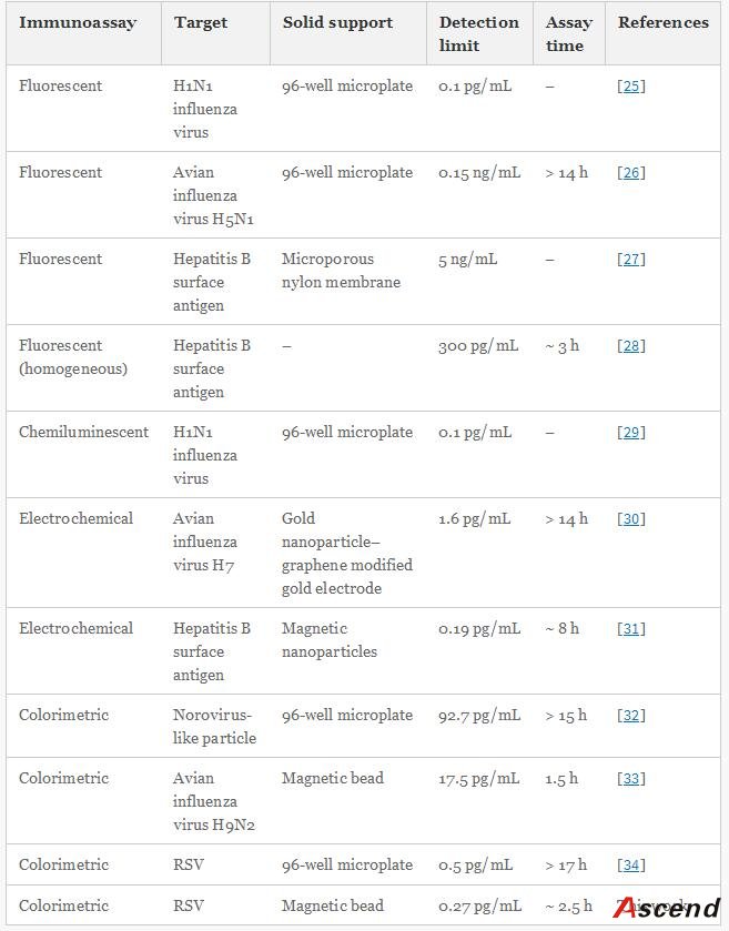
Furthermore, to improve the detection sensitivity of ELISA, incorporation of an additional signal amplification strategy is necessary. Up until now, some successful signal amplification systems utilizing enzymatic biocatalytic precipitation techniques or nanomaterials including liposomes and nanoparticles to load a large amount of tags have been proposed [14151617]. Among these strategies, the nanoparticle-based amplification has received a great deal of interest and caused a significant impact in immunoassay. In particular, gold nanoparticles (AuNPs) are very attractive materials for signal amplification in immunoassay due to their unique properties, such as good biological compatibility, high loading capacity, and stability [18]. Ambrosi et al. have fabricated an enhanced ELISA for the analysis of CA15–3 antigen using AuNPs as carriers of the signaling antibody anti-CA15–3-HRP (horseradish peroxidase) [19]. Though the assay holds higher sensitivity and shorter assay time when compared to conventional ELISA procedures, it suffers from complicated and expensive conjugation process of preparing HRP-linked antibodies. Lin et al. further increased HRP loading amount and thus reduced the detection limit and cost greatly by employing a combination of AuNPs and graphene oxide sheets as carriers [20]. However, this approach often requires multiple labels and multicycle operations for introducing nanoconjugates, resulting in the complicated, laborious, and time-consuming procedure.

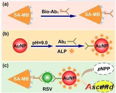
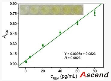
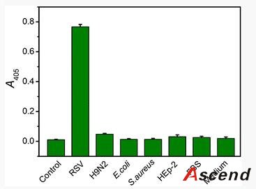
The work herein employs MBs as solid support and AuNPs as carrier to construct an enhanced immunoassay for the rapid, low-level detection of respiratory syncytial virus (RSV), which acts as a model target of pathogenic viruses. RSV, a single-stranded RNA virus of the paramyxocirus family, not only mediates serious lower respiratory tract illness in infants and toddlers which causes repeat infections throughout life, but also acts as a significant pathogen of the elderly and immune compromised patients [21]. Therefore, rapid, specific, and sensitive diagnosis of RSV is important to public-health monitoring.

Experimental

2.1 Materials and Apparatus

RSV strain was purchased from Guangzhou Bote Biological Technology Development Co., Ltd. (Guangzhou, China). The ELISA kit comprised a microtiter plate coated with anti-RSV, anti-RSV-HRP, hydrogen, TMB, 2 N sulfuric acid, a surfactant wash buffer, and RSV standard was purchased from Fengxiang Biotechnology Co., Ltd. (Shanghai, China). H9N2 avian influenza virus was obtained from Wuhan Institute of Virology, Chinese Academy of Sciences. Goat polyclonal anti-RSV biotin-conjugated antibody (Ab1, ab19986) and goat polyclonal anti-RSV antibody (Ab2, ab20745) were purchased from Abcam Inc. (Cambridge, MA). Streptavidin coated magnetic beads (MBs-SA), 4-nitrophenyl phosphate disodium salt hexahydrate (pNPP) and alkaline phosphatase (ALP) were obtained from Sigma-Aldrich (St. Louis, MO). HAuCl4·H2O was purchased from Sinopharm Group Chemical Regent Co., Ltd. (Shanghai, China). Bovine serum albumin (BSA) and trisodium citrate were obtained from Beijing Dingguo Changsheng Biotechnology Co. Ltd (Beijing, China). The phosphate buffer solution (PBS) consisted of 0.15 M NaCl, 2 mM KCl and 0.02 M phosphate buffer (pH 7.4). The diluted buffer of ALP consisted of 50% glycerol, 10 mM Tris, 5 mM MgCl2 and 0.1 mM ZnCl2 (pH 7.0). The detection buffer consisted of 50 mM Tris and 0.5 mM MgCl2 (pH 9.0). All reagents used in this experiment were of analytical grade without further purification, and Mili-Q purified water (18.2 MΩ) was used throughout the experiment.

The absorption spectra of AuNPs were recorded on a U-3010 spectrophotometer (Hitachi, Tokyo, Japan). The optical density (OD) at 405 nm was measured with a microplate reader (Biotek, Vermon, USA). JEM-2010 transmission electron microscopy (TEM) was used to identify the size and shape of gold nanoparticles. A Zetasizer Nano-ZS System (Malvern Inc.) was used for dynamic light scattering (DLS) measurements and zeta potential measurements.

2.2 Synthesis of AuNPs

AuNPs were prepared based on the classical citrate-reduction method with slight modifications [22]. Briefly, 2 mL of 1% (w/v) HAuCl4·H2O in 50 mL of ultrapure water was heated to reflux with vigorous stirring. Then, 1 mL of 5% (w/v) sodium citrate solution was rapidly introduced to react with HAuCl4, resulting in a color change of the mixture from pale yellow to gray, then purple, and finally to wine red. After 20 min, the heater was removed and the solution was left stirring and cooling down to room temperature. The product solution was stored at 4 °C for further use.

2.3 Preparation of MBs–Ab1 Complexes

Streptavidin-functionalized MBs were diluted to 0.1 mg/mL with PBS (pH 7.4). Then, 100 μL 100 μg/mL of biotinylated antibody (Bio-Ab1) was added to 1 mL of SA-MBs solution, and the mixed solution was incubated for 30 min at room temperature. Following that, the mixture was collected by applying an external magnetic field. Finally, the MBs–Ab1 was thoroughly washed three times with PBS (pH 7.4) and was resuspended into 1 mL of PBS (pH 7.4) containing 0.5% (w/v) BSA and stored at 4 °C for further usage.

2.4 Preparation of ALP–AuNPs–Ab2 Conjugates

The ALP–AuNPs–Ab2 conjugates were prepared by physical adsorption of Ab2 and ALP on AuNPs [23]. Briefly, the pH of AuNPs solution was adjusted to 9.0 with NaOH. Then, 100 μL of Ab2 (50 μg/mL) and 100 μL of ALP (180 μg/mL) were added to 1 mL of gold colloids. After gentle stirring for 2 h at room temperature, the mixture was further incubated overnight in the refrigerator to complete the reaction, which results in the adsorption of ALP and Ab2 on the surfaces of the AuNPs. Subsequently, the conjugates were centrifuged at 15,000 rpm for 20 min to remove the supernatant. The obtained precipitates (ALP–AuNPs–Ab2) were redispersed in 1 mL of PBS (pH 7.4) containing 0.5% (w/v) BSA and stored at 4 °C.

2.5 MBs-Based Immunoassay for RSV

MBs–Ab1 was used for fast target RSV collection and separation. 50 μL RSV with various concentrations and 50 μL of MBs–Ab1 suspension were successively injected into an Eppendorf tube. After incubation for 30 min at 37 °C with gentle shaking, the resulting mixture was magnetically collected and washed with PBS. Afterward, 100 μL of ALP–AuNPs–Ab2conjugates was added into the centrifuge tube and incubated for an additional 30 min under the same condition. The formed sandwich complexes between ALP–AuNPs–Ab2 and MBs–Ab1was separated and washed as the above protocol. Following that, the substrate solution (20 μL of 50 mM pNPP and 180 μL Tris–HCl) of ALP was introduced to the tube and shaken for 30 min for the color development. Subsequently, the reaction was stopped by adding 50 μL of 1 M NaOH and the optical density was recorded at 405 nm.

2.6 RSV Detection in Cell Lysate

RSV was propagated using HEp-2 human epithelial cells maintained in RPMI 1640 medium supplemented with 2% fetal bovine serum, penicillin (6 mg/mL) and streptomycin (10 mg/mL). Upon detectable cytopathic effect, RSV was harvested in serum-free medium followed by two freeze–thaws, after which the contents were collected and centrifuged at 3000g for 10 min. The virus concentration was determined by our method as described above and the standard ELISA method, respectively. The commercial ELISA kit was used in accordance with the manufacturer’s instructions.

Results and Discussion

3.1 Analytical Concept

The procedure of this immunoassay is described in Fig. 1. In this study, MB is employed as solid substrate to magnetically separate the immune complexes. Thus, as shown in Fig. 1a, the capture antibody (Ab1) is first specifically linked to the surfaces of MB via the fast and tight streptavidin–biotin interaction. For the immunoassay development, detection antibody (Ab2) and enzyme molecule (ALP) are immobilized on the citrate-protected spherical AuNP by physical adsorption to form ALP–AuNP–Ab2 conjugates (Fig. 1b). Subsequently, in the presence of RSV, the sandwiched immunocomplex between MB–Ab1 and ALP–AuNP–Ab2 can be formed following the illustration in Fig. 1c. The carried ALP molecules catalyze the dephosphorylation of non-colored pNPP to generate colored product pNP, which can be easily distinguished by the naked eye, making it suitable for detecting analytes in laboratories with fewer resources. In addition, since the amount of ALP is proportional to the concentration of RSV sandwiched through immunoreaction, the amount of RSV in the immunocomplex can be simply determined by measuring the optical intensity of the solution in a plate reader. Therefore, by employing MBs as solid substrate and AuNPs as enzyme carrier for signal amplification, this developed immunoassay would significantly improve the analytical performances with shorter time for the detection of RSV.
Open image in new window
Fig. 1

Schematic illustration of the preparation of a MBs–Ab1 complex, b ALP–AuNP–Ab2conjugate; and c principle of the sandwich colorimetric immunoassay for RSV

3.2 Characterization of the ALP–AuNPs–Ab2 Probe

AuNPs used in this immunoassay were prepared by reducing HAuCl4 with sodium citrate, thus they are surface coated by the citrate group. The as-prepared AuNPs are red in color, with a characteristic absorption peak at 518 nm, which is ascribed to the surface plasmon resonance of the AuNPs (Fig. 2a). Meanwhile, a TEM image shows that AuNPs are highly dispersed in aqueous solution and the average size is about 13 nm (Fig. 2b). After modification with ALP and antibody, the plasmon absorption peak of AuNPs shifted from 518 to 524 nm (Fig. 2a), suggesting that the biomolecules are successfully immobilized onto the AuNPs [24]. Furthermore, dynamic light scattering (DLS) was employed to analyze the hydrodynamic diameter of bare AuNPs and labeled AuNPs (Fig. 2c, d), which was increased from 17.5 ± 0.9 to 25.0 ± 0.1 nm after labeling, indicating the successful formation of the bioconjugates. Besides, the zeta potential measurements were also performed to characterize the change of surface charge after the formation of ALP–AuNPs–Ab2 conjugates. The surface potential of the bare AuNPs was −30.3 ± 0.7 mV because of the citrate groups capping, while it was neutralized to −9.9 ± 0.9 mV after conjugation, which could be ascribed to the effect of the positively charged amine groups (–NH3 +) in antibodies and enzymes. Taken together, the above results indicated the successful formation ALP–AuNPs–Ab2 conjugates.
Open image in new window
Fig. 2

Characterization of AuNPs and ALP–AuNPs–Ab2 conjugates. a UV–Vis absorption spectra of AuNPs (518 nm) and ALP–AuNPs–Ab2 (524 nm), b TEM images of AuNPs, cd DLS measurements of the average hydrodynamic sizes of bare AuNPs (c) and ALP–AuNPs–Ab2 conjugates (d)

To test the catalytic activity of ALP on the surfaces of AuNPs, ALP–AuNPs–Ab2 was reacted with pNPP, which is the substrate of ALP. As shown in Fig. 3, the AuNPs complex could convert the enzyme–substrate pNPP into a colored molecule pNP, which exhibited with a characteristic absorbance peak at about 405 nm. The results demonstrate that the ALP–AuNPs–Ab2 complex with catalytic activities could be further applied to the immunoassay for RSV.
Open image in new window
Fig. 3

UV-vis absorption spectra of the enzyme–substrate pNPP before (a) and after (b) reaction with the ALP–AuNPs–Ab2 conjugates. Fifty μL of diluted ALP–AuNPs–Ab2conjugates dispersion were reacted with 20 μL of 50 mM pNPP in PBS buffer. After incubation for 30 min, the mixture solution turned from colorless to bright yellow. The reaction was stopped by 1 M NaOH and the absorption spectra were recorded

3.3 Optimization of Experimental Conditions

To improve the sensitivity and reproducibility for RSV quantification, we have also optimized the experimental conditions, including the concentration of ALP and antibody, the pH for conjugation and the concentration of ALP–AuNP–Ab2 conjugates to be used in the immunoassay.

Citrate capped AuNPs are generally acknowledged to be relatively unstable and tend to aggregate irreversibly even in presence of a small amount of salt (NaCl), accompanying by a color change from red to blue. However, when AuNPs are functionalized with biomolecule, such as DNA or protein, the particles become much more stable and resistant to high concentrations of salt. Thus, to ensure the best gold surface coverage, the gold aggregation test was preliminarily performed to judge the optimal pH value to use for the anti-RSV antibody and ALP conjugation because the conjugation process is influenced greatly by the pH which determines the charges and stability of the protein. In this work, the optimal conditions for the preparation of ALP–AuNP–Ab2 conjugates were determined by measuring the difference between the absorbance at 520 and at 680 nm and plotting it against the different pH values (Fig. 4a, b). As can be seen, the highest level of AuNPs surface coverage by the anti-RSV antibody is achieved when the complex prepared at pH 9.0, while the optimal pH at which gives the highest absorbance difference is found around 7.0–9.0. Taken together, the pH of AuNPs solution was adjusted to pH 9.0 for the conjugation of anti-RSV antibody and ALP to AuNPs. Similarly, the antibody concentration that prevents AuNPs aggregation was also determined by the gold aggregation test. The minimum antibody concentration for conjugation is 10 μg for 1 mL of AuNPs (Fig. 4c). Meanwhile, with the increasing concentration of antibody, the spectrum of AuNPs–anti-RSV conjugate solutions became more and more similar to that of bare AuNPs, indicating that the surface of AuNPs is fully covered by antibody molecules (Fig. 4d). In addition, it was found 36.5 μg/mL of ALP molecules was necessary to completely cover the AuNPs surface (Fig. 4e). Based on the principle of low cost and high sensitivity, 5 μg anti-RSV antibody and 18 μg ALP was adopted for the preparation of ALP–AuNPs–Ab2conjugates in the subsequent study.
Open image in new window
Fig. 4

Optimization of the conditions for the preparation of the ALP–AuNPs–Ab2 conjugates (Influence of pH in the conjugation of anti-RSV antibody (a) and ALP (b) to AuNPs, (c) Effect of anti-RSV antibody concentration on the conjugation and the corresponding spectra (d), (e) Effect of ALP concentration on the conjugation.) and the concentration of the ALP–AuNPs–Ab2 conjugates used for RSV detection in developed immunoassay (f)

Furthermore, the concentration of the ALP–AuNPs–Ab2 conjugates to be used in the immunoassay was also optimized (Fig. 4f). Various dilution coefficients of 1:2, 1:5, 1:20, 1:50 and 1:100 were analyzed. As the concentration of ALP–AuNPs–Ab2 increased, the value of optical density increased accordingly. However, when a high complex concentration (1:2) was employed, the negative control (without RSV) exhibited a high nonspecific signal, which significantly lowered the signal-to-noise ratio. Taken together, the optimal dilution, with a good balance between low nonspecific signal and high sensitivity, was 1:20.

3.4 Analytical Characteristics for RSV Detection

Analytical performance characteristics of the method were determined by measuring the absorbance at 405 nm with varying concentration of RSV. Figure 5 shows the color signal and optical response produced by our method for the detection of RSV. There was a good linear relationship between the optical signal and the concentration of RSV in the range of 0.5–80 pg/mL, and the regression equation was Y = 0.0096 x + 0.0523 (the correlation coefficient R = 0.9923), where Y is the absorption intensity, and x is the concentration of RSV. The limit of detection (LOD, 3σ/k) for this immunoassay is 0.27 pg/mL, which is over sevenfold lower than that of commercially available RSV ELISA kit (2 pg/mL). Furthermore, using MBs as solid substrate to magnetically separate the immune complexes, which eliminates the multiple washing steps in conventional ELISA, the total assay time was decreased to less than 2.5 h without any pretreatment. Therefore, by taking advantage of the magnetic separation and enrichment of MBs and signal amplification of AuNPs carrier, our strategy provide an enhanced immunoassay for fast and sensitive detection of viral pathogens in clinical diagnosis. Furthermore, the performance of the novel method has been compared with other immunoassays for viral pathogens. Characteristics such as the detection limit and assay time are summarized for all of them in Table 1. As can be observed, the detection limit of the developed immunoassay is competitive with the most sensitive viral detection techniques and the assay time by employing MBs as solid support is much lower than those of other microplate-based immunoassays. The reason for sensitivity enhancement is attributed to the fact that AuNPs can carry multi-ALP molecules, and they gave a higher catalytic efficiency, which therefore, amplify the enzymatic signal. On the other hand, the use of MBs not only reduces the immunoreaction time but also amplifies the signal due to an increase in the surface area and the magnetic enrichment effect.
Open image in new window
Fig. 5

MBs-based sandwich immunoassay performed for RSV. The inset shows the corresponding photograph. The concentrations of RSV were 0.5, 5, 20, 40, 60, 80 pg/mL, respectively. ALP–AuNPs–Ab2, 1:20; detection buffer, 5 mM pNPP, 50 mM Tris–HCl, 0.5 mM MgCl2, pH 9.0. The error bars represent the standard deviation of three measurements

Table 1

Brief summary of the results reported on several immunoassays for the detection of viral pathogen

The specificity of the immunoassay played an important role in analyzing pathogens in complex biological samples in situ without any pretreatment. Therefore, to assess the specificity of the proposed immunoassay for RSV detection, other antigens such as H9N2 influenza virus, Gram-negative bacteria Escherichia coli (E. coli), Gram-positive bacteria Staphylococcus aureus (S. aureus), human epithelial type 2 (HEp-2) cells, fetal bovine serum (FBS), and RPMI-1640 medium were employed to investigate following the same procedure. As indicated clearly in Fig. 6, obviously higher optical intensity can be observed with the target virus than that of other components, demonstrating the high specificity of the developed immunoassay. In addition, the reproducibility of the immunoassay for RSV was investigated with intra-assay and interassay precision. The intra-assay precision was performed by analyzing one RSV level for five repetitive measurements, while the interassay precision was carried out by determining one RSV level with five times independently. The intra- and interassay variation coefficients obtained from 20 pg/mL RSV were 5.2 and 7.1%, respectively, indicating that the immunoassay possessed acceptable reproducibility.
Open image in new window
Fig. 6

Specificity test of the immunoassay for RSV. The concentration of RSV is 80 pg/mL, while H9N2 is 0.5 ng/mL; the number of E. coli and S. aureus is 8 × 109 CFU/mL; the number of HEp-2 is 106/mL. The error bars represent the standard deviation of three measurements

Finally, to evaluate the capability of our method in complex samples, RSV-infected cell lysates were analyzed using our method and the standard ELISA for the quantification of RSV. Table 2shows the RSV concentrations obtained via each method in the different batches of cell lysates. It is found that our method gives results comparable to those of the ELISA, indicating that the proposed method is reliable and it is also suitable for the quantification of RSV in biological samples.
Table 2

The amount of RSV present in different batches of cell lysates samples using the proposed immunoassay and the standard ELISA

Batches

Proposed immunoassay (pg/mL)

ELISA (pg/mL)

1

45.01 ± 1.15

45.73 ± 0.66

2

16.84 ± 1.02

17.11 ± 0.83

3

33.15 ± 0.72

32.89 ± 0.64

Conclusions

In summary, we have demonstrated the feasibility of a sandwich colorimetric immunoassay for the determination of RSV using MBs as capture substrate and AuNPs as carrier for signal amplification. Compared with conventional ELISA, this method was further improved with enhanced sensitivity, decreased analysis time and rapid separation process. Moreover, it can be easily extended for the detection of other viral pathogens since the biofunctionalization process is very simple and generally applicable. In the future, to further improve the analytical performances and reduce the analysis time, efforts to construct microfluidic device that allow high throughput processing with minimal time and material/reagent consumption by integrating MBs and AuNPs are ongoing.

Notes

Acknowledgements

This work was supported by the National Natural Science Foundation of China (NSFC, No. 21535006), the National Basic Research Program of China (973 Program, No. 2011CB933600), and the Doctoral Scientific Research Foundation (SWU116058).

Compliance with Ethical Standards

Conflict of interest

The author(s) declare that they have no competing interests.

纳米颗粒:www.ex-dna.com

References

  1. 1.
    Granger JH, Schlotter NE, Crawford AC, Porter MD. Prospects for point-of-care pathogen diagnostics using surface-enhanced Raman scattering (SERS). Chem Soc Rev. 2016;45:3865–82.CrossRefGoogle Scholar
  2. 2.
    Vaculovicova M, Michalek P, Krizkova S, Macka M, Adam V. Nanotechnology-based analytical approaches for detection of viruses. Anal Methods. 2017;9:2375–91.CrossRefGoogle Scholar
  3. 3.
    Chen CM, Martinez AW, Gong J, Mace CR, Phillips ST, Carrilho E, Mirica KA, Whitesides GM. Paper-based ELISA. Angew Chem Int Ed. 2010;49:4771–4.CrossRefGoogle Scholar
  4. 4.
    Hsing I, Xu Y, Zhao W. Micro- and nano-magnetic particles for applications in biosensing. Electroanalysis. 2007;19:755–68.CrossRefGoogle Scholar
  5. 5.
    Kim YS, Jurng J. A simple colorimetric assay for the detection of metal ions based on peroxidase-like activity of magnetic nanoparticles. Sens Actuat B Chem. 2013;176:253–7.CrossRefGoogle Scholar
  6. 6.
    Wei H, Wang E. Fe3O4 magnetic nanoparticles as peroxidase mimetics and their applications in H2O2 and glucose detection. Anal Chem. 2008;80:2250–4.CrossRefGoogle Scholar
  7. 7.
    Yang YC, Wang YT, Tseng WL. Amplified peroxidase-like activity in iron oxide nanoparticles using adenosine monophosphate: application to urinary protein sensing. ACS Appl Mater Interfaces. 2017;9:10069–77.CrossRefGoogle Scholar
  8. 8.
    Park KS, Kim MI, Cho DY, Park HG. Label-free colorimetric detection of nucleic acid based on target-induced shielding against the peroxidase-mimicking activity of magnetic nanoparticles. Small. 2011;7:1521–5.CrossRefGoogle Scholar
  9. 9.
    Wu J, Wei X, Gan J, Huang L, Shen T, Lou J, Liu B, Zhang JX, Qian K. Multifunctional magnetic particles for combined circulating tumor cells isolation and cellular metabolism detection. Adv Funct Mater. 2016;26:4016–25.CrossRefGoogle Scholar
  10. 10.
    Adams NM, Bordelon H, Wang KK, Albert LE, Wright DW, Haselton FR. Comparison of three magnetic bead surface functionalities for RNA extraction and detection. ACS Appl Mater Interfaces. 2015;7:6062–9.CrossRefGoogle Scholar
  11. 11.
    Heun Y, Hildebrand S, Heidsieck A, Gleich B, Anton M, Pircher J, Ribeiro A, Mykhaylyk O, Eberbeck D, Wenzel D, Pfeifer A, Woernle M, Krötz F, Pohl U, Mannell H. Targeting of magnetic nanoparticle-coated microbubbles to the vascular wall empowers site-specific lentiviral gene delivery in vivo. Theranostics. 2017;7:295–307.CrossRefGoogle Scholar
  12. 12.
    Guo X, Li W, Luo L, Wang Z, Li Q, Kong F, Zhang H, Yang J, Zhu C, Du Y, You J. External magnetic field-enhanced chemo-photothermal combination tumor therapy via iron oxide nanoparticles. ACS Appl Mater Interfaces. 2017;9:16581–93.CrossRefGoogle Scholar
  13. 13.
    Tennico YH, Hutanu D, Koesdjojo MT, Bartel CM, Remcho VT. On-chip aptamer-based sandwich assay for thrombin detection employing magnetic beads and quantum dots. Anal Chem. 2010;82:5591–7.CrossRefGoogle Scholar
  14. 14.
    Akama K, Shirai K, Suzuki S. Droplet-free digital enzyme-linked immunosorbent assay based on a tyramide signal amplification system. Anal Chem. 2016;88:7123–9.CrossRefGoogle Scholar
  15. 15.
    Ye H, Yang K, Tao J, Liu Y, Zhang Q, Habibi S, Nie Z, Xia X. An enzyme-free signal amplification technique for ultrasensitive colorimetric assay of disease biomarkers. ACS Nano. 2017;11:2052–9.CrossRefGoogle Scholar
  16. 16.
    Wei T, Du D, Zhu MJ, Lin Y, Dai Z. An improved ultrasensitive enzyme-linked immunosorbent assay using hydrangea-like antibody-enzyme-inorganic three-in-one nanocomposites. ACS Appl Mater Interfaces. 2016;8:6329–35.CrossRefGoogle Scholar
  17. 17.
    Lin C, Guo Y, Zhao M, Sun M, Luo F, Guo L, Qiu B, Lin Z, Chen G. Highly sensitive colorimetric immunosensor for influenza virus H5N1 based on enzyme-encapsulated liposome. Anal Chim Acta. 2017;963:112–8.CrossRefGoogle Scholar
  18. 18.
    Zheng W, Jiang X. Integration of nanomaterials for colorimetric immunoassays with improved performance: a functional perspective. Analyst. 2016;141:1196–208.CrossRefGoogle Scholar
  19. 19.
    Ambrosi A, Airò F, Merkoçi A. Enhanced gold nanoparticle based ELISA for a breast cancer biomarker. Anal Chem. 2010;82:1151–6.CrossRefGoogle Scholar
  20. 20.
    Lin H, Liu Y, Huo J, Zhang A, Pan Y, Bai H, Jiao Z, Fang T, Wang X, Cai Y, Wang Q, Zhang Y, Qian X. Modified enzyme-linked immunosorbent assay strategy using graphene oxide sheets and gold nanoparticles functionalized with different antibody types. Anal Chem. 2013;85:6228–32.CrossRefGoogle Scholar
  21. 21.
    Griffiths C, Drews SJ, Marchant DJ. Respiratory syncytial virus: infection, detection, and new options for prevention and treatment. Clin Microbiol Rev. 2017;30:277–319.CrossRefGoogle Scholar
  22. 22.
    Wang J, Wu ZL, Zhang HZ, Li YF, Huang CZ. Selective colorimetric analysis of spermine based on the cross-linking aggregation of gold nanoparticles chain assembly. Talanta. 2017;167:193–200.CrossRefGoogle Scholar
  23. 23.
    Li YS, Zhou Y, Meng XY, Zhang YY, Liu JQ, Zhang Y, Wang NN, Hu P, Lu SY, Ren HL, Liu ZS. Enzyme–antibody dual labeled gold nanoparticles probe for ultrasensitive detection of & #x03BA;-casein in bovine milk samples. Biosens Bioelectron. 2014;61:241–4.CrossRefGoogle Scholar
  24. 24.
    Kumar S, Aaron J, Sokolv K. Directional conjugation of antibodies to nanoparticles for synthesis of multiplexed optical contrast agents with both delivery and targeting moieties. Nat Protoc. 2008;3:314–20.CrossRefGoogle Scholar
  25. 25.
    Li Y, Hong M, Qiu B, Lin Z, Chen Y, Cai Z, Chen G. Highly sensitive fluorescent immunosensor for detection of influenza virus based on Ag autocatalysis. Biosens Bioelectron. 2014;54:358–64.CrossRefGoogle Scholar
  26. 26.
    Chen L, Sheng Z, Zhang A, Guo X, Li J, Han H, Jin M. Quantum-dots-based fluoroimmunoassay for the rapid and sensitive detection of avian influenza virus subtype H5N1. Luminescence. 2010;25:419–23.CrossRefGoogle Scholar
  27. 27.
    Liu Y, Brandon R, Cate M, Peng X, Stony R, Johnson M. Detection of pathogens using luminescent CdSe/ZnS dendron nanocrystals and a porous membrane immunofilter. Anal Chem. 2007;79:8796–802.CrossRefGoogle Scholar
  28. 28.
    Zeng Q, Li Q, Ji W, Bin X, Song J. Highly sensitive homogeneous immunoassay based on construction of silver triangular nanoplates-quantum dots FRET system. Sci Rep. 2016;6:26534.CrossRefGoogle Scholar
  29. 29.
    Li Y, Hong M, Qiu B, Lin Z, Cai Z, Chen Y, Chen G. A highly sensitive chemiluminescent metalloimmunoassay for H1N1 influenza virus detection based on a silver nanoparticle label. Chem Commun. 2013;49:10563–5.CrossRefGoogle Scholar
  30. 30.
    Huang J, Xie Z, Xie Z, Luo S, Xie L, Huang L, Fan Q, Zhang Y, Wang S, Zeng T. Silver nanoparticles coated graphene electrochemical sensor for the ultrasensitive analysis of avian influenza virus H7. Anal Chim Acta. 2016;913:121–7.CrossRefGoogle Scholar
  31. 31.
    Alizadeh N, Hallaj R, Salimi A. A highly sensitive electrochemical immunosensor for hepatitis B virus surface antigen detection based on Hemin/G-quadruplex horseradish peroxidase-mimicking DNAzyme-signal amplification. Biosens Bioelectron. 2017;94:184–92.CrossRefGoogle Scholar
  32. 32.
    Ahmed SR, Takemeura K, Li TC, Kitamoto N, Tanaka T, Suzuki T, Park EY. Size-controlled preparation of peroxidase-like graphene-gold nanoparticle hybrids for the visible detection of norovirus-like particles. Biosens Bioelectron. 2017;87:558–65.CrossRefGoogle Scholar
  33. 33.
    Zhou CH, Zhao JY, Pang DW, Zhang ZL. Enzyme-induced metallization as a signal amplification strategy for highly sensitive colorimetric detection of avian influenza virus particles. Anal Chem. 2014;86:2752–9.CrossRefGoogle Scholar
  34. 34.
    Zhan L, Wu WB, Yang XX, Huang CZ. Gold nanoparticle-based enhanced ELISA for respiratory syncytial virus. New J Chem. 2014;38:2935–40.CrossRefGoogle Scholar